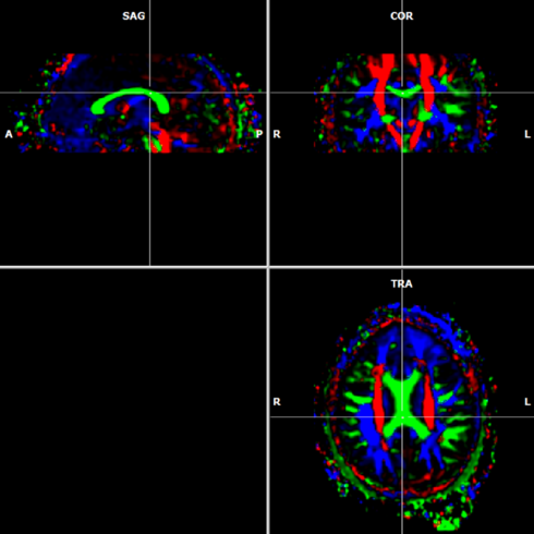
FA color coding is incorrect. Why? Because co-ordinate system of scanner and BrainVoyager is different. This is not a BV problem, since co-ordinate systems are not standardized.
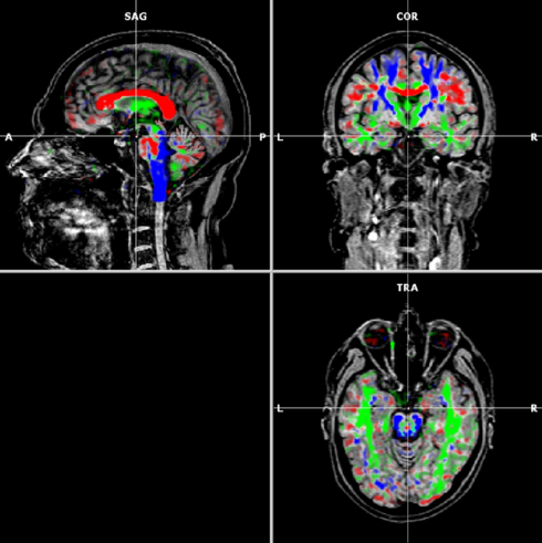
Normal color coding:
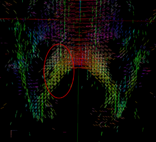
An example of abnormal color coding:
| Example: Original color | Proposed change |
| Ant-Post = blue | X: direction = Inf-Sup |
| Inf-Sup = red | Y: direction = Left-Right |
| Left-Right = green | Z: direction = Ant-Post |
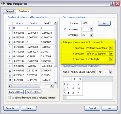
Procedure to correct:
go to DTI -> Diffusion Weighted Data Analysis -> VDW Properties ->
Gradients. Change x, y and z to what you think is correct. This is a mathematical
operation and has nothing to do with orientation in the brain. Next, click the OK
button.
Re-calculate the tensors by clicking the Estimate button. In the Calculations tab, create the color coded FA maps and check them for the right colors.
Then, Go to DTI -> Tensor visualisation and check for tensor directions. I always use the splenium of the corpus callosum to check for correct orientation.
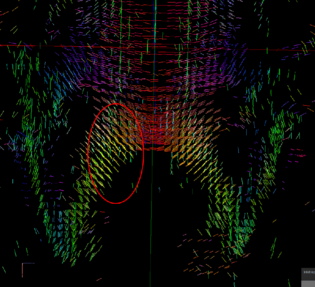
(a) incorrect tensors |
(b) correct tensors |