EPI artifacts
- Details
- Category: Pre-Processing
- Last Updated: 09 April 2018
- Published: 09 April 2018
- Hits: 5739
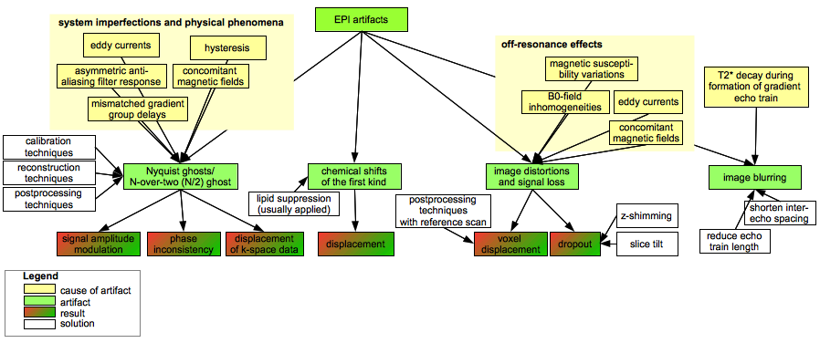
Please find below a summary of echo planar imaging (EPI) artifacts, based on Chapter 16.1.4 of Bernstein et al (2004) Handbook of MRI sequences.
Related articles on the support site
Checking DMR data quality - f.e. eddy currents
Fieldmap based EPI distortion correction - anatabacus plugin
More information
For more information about and illustrative examples of EPI artifacts, see for example
- Hornak's book "The basis of MRI"; the chapter about artifacts: http://www.cis.rit.edu/htbooks/mri/chap-11/chap-11.htm#11.3
- JEMRIS MRI simulation framework: generation and illustration of some artifacts
- Signal dropout: N. Weiskopf et al.(2006) Optimal EPI parameters for reduction of susceptibility-induced BOLD sensitivity losses: A whole-brain analysis at 3 T and 1.5 T. NeuroImage 33 (2006) 493–504. And a possible cure at 7T: Brunheim et al (2014) Variable slice-thickness (VAST) EPI for the reduction of signal voids in GE-EPI at 7 Tesla. Presented at the Human Brain Mapping conference in Hamburg.