The optimal transformation to undistorted images is completely determined by the image registration procedure itself; it does not use any prior information.
To evaluate the results of the algorithm, one can perform visual inspection, quantification by numbers, check the results with a B0 fieldmap or simulations.
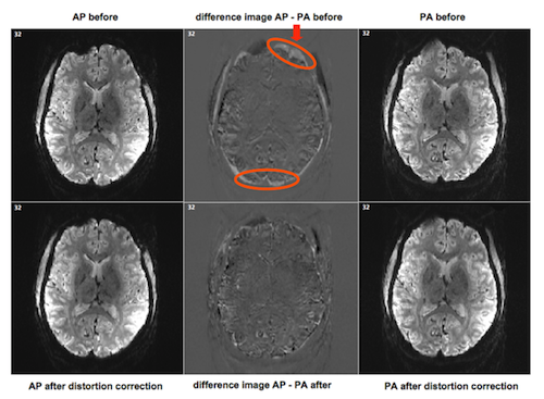
The idea is to create a difference image of the opposite phase encoded images before and after running unwarping via COPE. Via the "Evaluation" tab in the anatabacus plugin it is possible to subtract FMR files from each other. If the images are more similar after distortion correction, the edges at top and bottom (AP-PA case) or left and right (LR-RL case) of the brain should be less wide, which shows that the expansion and compression due to the susceptibility artifact have been undone.
To compare the difference images properly, set contrast and brightness at similar values, for example 50-50 (via BrainVoyager menu > Options > Contrast and Brightness..).
For checking specific regions, it might be good to reduce the number of columns and rows shown of the FMR to just one or a few and zoom in. Look if structures like the ventricles and obvious sulci are matching after being undistorted. There can be quite a difference before and after the correction:
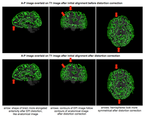
It is also possible to perform initial alignment to an anatomical T1 weighted image before and after EPI distortion correction. After distortion correction, the contours of the EPI image should follow the contours of the anatomical image more accurately.
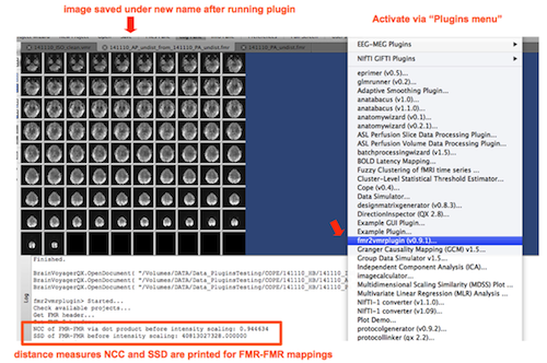
A distance measure can be used to give single number that expresses the similarity between the two opposite phase encoded images; the distance between the original AP and PA (or LR and RL) images should be larger than the distance between those images after applying distortion correction.
With the FMR->VMR mapping and the FMR->FMR mapping in the fmr2vmrplugin it is possible to obtain distance measures (see figure below). For download, see the section Plugins on the BrainVoyager support site.
If a B0 fieldmap is available, it should be possible to compare the results from EPI distortion correction via image registration of opposite phase encoded images with undistortion by a B0 fieldmap (although, in a study with two 3T and two 7T datasets, the results obtained by fieldmap gave lower similarity after correction than with image-registration based undistortion via TOPUP and HySCO, see Fritz et al (2014)[1]).
In theory, it should also be possible to check with simulated B0 maps and simulated EPI data whether the distortions were corrected properly. MRI simulators are for example Jemris (Juelich) and POSSUM (Oxford).
Latest update of this page: 11 February 2015